How a Cardiologist Reads an American Shorthair’s Scan
The thought of your cherished American Shorthair needing a heart scan can bring a wave of anxiety. It’s a specialized procedure that often raises more questions than answers initially, leaving you wondering exactly what the veterinary cardiologist is looking for and what all those technical terms mean for your beloved feline. This comprehensive guide will walk you through the intricate process of an American Shorthair’s heart scan, demystifying how a cardiologist reads the results and what those findings might indicate about your cat's health.
Facing the Unknown: Why Your American Shorthair Needs a Specialized Heart Scan
It’s a tough moment when your regular vet suggests a referral to a veterinary cardiologist. Maybe they heard a subtle heart murmur during a routine check-up, or perhaps your American Shorthair is showing some very mild, almost imperceptible changes at home that raised a flag. The natural stoicism of the American Shorthair means they often hide illness incredibly well, so by the time obvious symptoms appear, a condition might be quite advanced. This breed, while generally robust, isn't immune to certain health challenges, and one of the most significant concerns for American Shorthair owners, especially breeders, is hypertrophic cardiomyopathy, or HCM.
HCM is the most common heart disease in cats, characterized by a thickening of the heart muscle, particularly the left ventricle. This thickening makes it harder for the heart to pump blood effectively, and over time, it can lead to serious complications like congestive heart failure, blood clots, or even sudden death. For a long time, breeders and owners have grappled with this silent threat. While the American Shorthair is celebrated for its genetic soundness, HCM Is the One Inherited Disease the American Shorthair Kept. This isn't a reflection on the breed's overall health but rather a reminder that even the hardiest lines can carry genetic predispositions. Therefore, understanding Why an American Shorthair Still Needs a Yearly Heart Scan is paramount, as early detection through a specialist's eye can make all the difference in managing the condition and ensuring a good quality of life.
The reason a full cardiac workup goes beyond listening to a murmur is that not all murmurs indicate significant heart disease, and conversely, some cats with severe HCM may not have an audible murmur at all. This is where the echocardiogram, essentially an ultrasound of the heart, becomes an indispensable diagnostic tool. It allows the cardiologist to visualize the heart's structure and function in real-time, offering a level of detail no stethoscope or X-ray can provide. It's a critical step, particularly because Why a Gene Test Cannot Clear an American Shorthair's Heart means genetic testing, while available for some breeds, isn't a definitive "all-clear" for the American Shorthair.
Behind the Scenes: What Happens During an Echocardiogram for Your American Shorthair
When you bring your American Shorthair in for an echocardiogram, it's a very different experience than a typical vet visit. You'll likely be at a specialist clinic, possibly in a quiet, dedicated room designed to minimize stress for your cat. The procedure itself is non-invasive and generally well-tolerated. Your cat will usually lie comfortably on a special table, often with a cut-out section, allowing the cardiologist to easily position the ultrasound probe against their chest. Some cats might need a small patch of fur shaved to ensure good contact and clear imaging, but often, the fur is simply parted and alcohol gel applied.
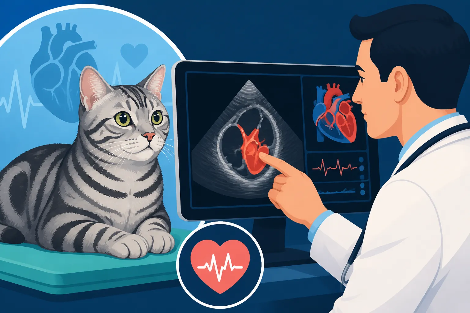
One of the most reassuring aspects for many owners is that sedation is usually not required for an echocardiogram in cats, especially for a generally calm and cooperative breed like the American Shorthair. Cardiologists are highly skilled at keeping cats relaxed throughout the approximately 20-40 minute procedure. They use gentle handling techniques and sometimes have veterinary technicians whose sole job is to comfort and stabilize your cat. The environment is typically kept calm and quiet, which helps reduce any anxiety your cat might feel. The room will have an ultrasound machine with a monitor, and the cardiologist will apply a water-soluble gel to the transducer (the probe) to facilitate sound wave transmission and get the clearest images.
During the scan, the cardiologist will systematically capture various views of your cat's heart. They'll move the probe across the chest, looking at different angles and depths. The ultrasound machine then translates these sound waves into real-time images on the screen. It's truly incredible to watch your cat's heart beating live, and the cardiologist will be meticulously measuring, noting, and interpreting what they see. This comprehensive approach ensures that every aspect of the heart’s anatomy and function is evaluated, providing a complete picture for an accurate diagnosis. It's this detailed, live imaging that makes the echocardiogram the gold standard for diagnosing feline heart conditions, leaving no room for guesswork.
The Cardiologist's Eye: Deciphering the Nuances of Your American Shorthair's Heart
This is where the magic, or rather, the advanced medical expertise, really happens. The cardiologist isn't just looking at a pretty picture of a beating heart; they are performing a detailed anatomical and functional analysis. They use several modes of ultrasound imaging to gather crucial data, comparing their findings against established normal values for cats, and sometimes specifically for robust breeds like the American Shorthair.
B-Mode and M-Mode Imaging: Structural Assessment
The primary components of an echocardiogram are typically 2D (B-mode) and M-mode imaging.
- B-mode (Brightness Mode or 2D): This provides real-time, two-dimensional cross-sectional images of the heart. The cardiologist uses this to assess the overall shape, size, and movement of the heart chambers, valves, and major blood vessels. They'll look at the thickness of the ventricular walls, particularly the left ventricle, which is the main pumping chamber. In HCM, this wall will appear abnormally thick. They'll also check the size of the atria, especially the left atrium, which can enlarge as the heart works harder against the thickened muscle. A key measurement here is the left atrium to aorta ratio (LA:Ao), where an elevated ratio can indicate increased pressure and volume within the left atrium, a common sign of advanced HCM. They also carefully examine the papillary muscles, small muscle structures inside the ventricles that anchor the heart valves, as these can also become hypertrophied in American Shorthairs with HCM.
- M-mode (Motion Mode): This provides a one-dimensional, "ice pick" view of the heart's motion over time. It's incredibly precise for measuring wall thickness, chamber dimensions, and the movement of heart valves during the cardiac cycle. The cardiologist will measure the thickness of the left ventricular free wall and septum during both systole (contraction) and diastole (relaxation). These precise measurements are critical for diagnosing HCM. They'll also use M-mode to calculate parameters like fractional shortening (FS), which is an indicator of the heart's systolic function, or how effectively it's pumping blood out. A reduced fractional shortening can signal impaired pump function.
Doppler Imaging: Assessing Blood Flow Dynamics
Beyond structure, understanding blood flow is crucial. Doppler echocardiography uses sound waves to evaluate the speed and direction of blood flow within the heart and great vessels.
- Color Flow Doppler: This technique superimposes color onto the 2D image, with different colors indicating blood flow direction and velocity. It's invaluable for detecting abnormalities like regurgitation (blood flowing backward through a valve) or turbulent flow. For instance, if the mitral valve isn't closing properly due to the thickened muscle pulling on it (a condition called Systolic Anterior Motion of the Mitral Valve, or SAM), color Doppler will show blood leaking backward into the left atrium, known as mitral regurgitation. This is a common finding in many cats with HCM and can further strain the heart.
- Pulsed Wave and Continuous Wave Doppler: These advanced Doppler modes allow the cardiologist to precisely measure the velocity of blood flow at specific points. They are particularly interested in the outflow tract of the left ventricle, the area where blood exits the heart. In some cases of HCM, the thickened heart muscle can create an obstruction here, leading to very high-velocity blood flow through a narrowed opening. This is called Left Ventricular Outflow Tract Obstruction (LVOTO) and is a significant finding that influences treatment decisions.
By combining all these imaging modes, the cardiologist builds a comprehensive picture of your American Shorthair's heart health. They are looking for the subtle and not-so-subtle changes that might indicate disease, especially because The Age an American Shorthair's Heart Starts to Thicken can vary, and early detection is key. For example, some American Shorthairs might not show any external signs, yet their hearts are undergoing significant changes internally, reinforcing the importance of regular screening for at-risk individuals or those with a family history.
Beyond the Numbers: Understanding the Diagnosis and What Comes Next for Your American Shorthair
Once the cardiologist has meticulously gathered all the images and measurements, they will interpret the findings to provide a diagnosis and prognosis. This conversation can be incredibly daunting, but understanding the categories helps.
Interpreting the Results
Generally, results fall into a few key categories:
- Normal: This is the best news! It means all measurements and observations fall within the healthy range. Even with a normal scan, if your American Shorthair is part of a breeding program or has specific risk factors, annual screening might still be recommended.
- Equivocal or Borderline HCM: This can be a tricky category. It means there are some minor changes, perhaps slight wall thickening that doesn't definitively meet the criteria for HCM but is enough to warrant concern. It could be an early stage of the disease, or a variant of normal. In these cases, the cardiologist will likely recommend a follow-up scan in 6-12 months to monitor for progression. This highlights the importance of regular check-ups, even if a cat is showing no outward What an American Shorthair Shows Before Its Heart Fails.
- Mild, Moderate, or Severe HCM: If the criteria for HCM are met, the cardiologist will grade the severity.
- Mild HCM usually means definite thickening is present but the heart's function is still largely normal, and there are typically no clinical signs.
- Moderate HCM indicates more significant thickening and possibly some left atrial enlargement or mild obstruction. Your cat might still be asymptomatic, but the risk of complications increases.
- Severe HCM means pronounced wall thickening, often with significant left atrial enlargement, potential severe outflow tract obstruction, and sometimes evidence of early congestive heart failure. Cats with severe HCM are at higher risk for clinical signs and complications.
The prognosis varies widely depending on the severity of the disease and how quickly it progresses. Some cats with mild HCM can live for years without developing significant problems, while others with more aggressive forms may have a shorter lifespan.
The Path Forward: Management and Monitoring
Regardless of the diagnosis, the cardiologist will develop a tailored plan for your American Shorthair.
- For Normal and Borderline Cases: The main recommendation is often continued monitoring. This usually involves regular follow-up echocardiograms to detect any changes over time. Your vet might also suggest keeping a close eye on your cat's activity level, appetite, and breathing at home.
- For Diagnosed HCM Cases: Management typically focuses on slowing disease progression and preventing complications.
- Medication: This is often prescribed to reduce heart rate, improve relaxation of the heart muscle, and prevent blood clot formation. Common medications include beta-blockers (like atenolol), calcium channel blockers (like diltiazem), ACE inhibitors, and in some cases, blood thinners (like clopidogrel). Which Drugs Slow an American Shorthair's Heart Disease will depend entirely on the specific findings and severity.
- Dietary Modifications: While not a cure, a low-sodium diet might be recommended, especially if there are signs of heart failure, to help manage fluid retention.
- Close Monitoring: Regular veterinary check-ups are essential, often every 3-6 months, including repeat echocardiograms to assess the effectiveness of medication and monitor for any changes. Owners are usually advised to monitor for signs like increased breathing rate, lethargy, or coughing, which could indicate progression to heart failure. How a Vet Manages an American Shorthair With HCM is a dynamic process, adjusting treatment as needed.
It's important to remember that these specialized scans and ongoing treatments can be costly. This is where pet insurance, like plans offered by Healthy Paws, can be an absolute lifesaver. Having a good policy in place before any diagnosis can significantly ease the financial burden of managing a chronic condition like HCM, allowing you to focus on your cat's well-being without added stress. Understanding Whether Insurance Pays for an American Shorthair's Echo is a good question to ask your provider well in advance, as specialized diagnostics are often covered under comprehensive plans.
Receiving a diagnosis of HCM can be overwhelming, but knowledge is power. Understanding how a cardiologist meticulously reads an American Shorthair's heart scan empowers you to be an informed advocate for your cat's health. With careful monitoring and appropriate management, many American Shorthairs with HCM can still enjoy a good quality of life for many years.
| Echocardiogram Parameter | Normal American Shorthair (Approximate) | Findings Indicative of HCM | Why it Matters |
|---|---|---|---|
| Left Ventricular Wall (Diastole) | < 5.5 mm | > 6 mm (especially septal & free wall) | Primary diagnostic criterion for HCM; indicates thickened heart muscle. |
| Left Atrial to Aorta (LA:Ao) Ratio | < 1.5 | > 1.6 – 1.7 (and often much higher) | Indicates left atrial enlargement due to increased pressure/volume in the left atrium. |
| Papillary Muscle Size | Normal | Hypertrophied, enlarged | Thickening of these muscles also contributes to overall heart muscle thickening in HCM. |
| Systolic Anterior Motion (SAM) | Absent | Present (mitral valve leaflet moves into outflow tract) | Obstructs blood flow from the left ventricle, causing turbulence and pressure gradient. |
| Left Ventricular Outflow Tract (LVOT) Velocity | Normal (< 2 m/s) | Elevated (> 2 m/s, often > 4 m/s) | Indicates dynamic obstruction of blood leaving the heart, leading to increased workload. |
| Fractional Shortening (FS) | 30-50% | Can be normal, decreased, or even increased (in early stages due to hypercontractility) | Measures pumping efficiency; later stages of HCM can show decreased function. |
Note: These values are approximations and can vary slightly based on individual cat size, specific views, and the cardiologist's discretion. A definitive diagnosis requires a full assessment by a board-certified veterinary cardiologist.
Disclaimer
This article is intended for informational purposes only and does not constitute medical advice. Always consult with a qualified veterinary professional for any health concerns or before making any decisions related to your American Shorthair's care.